Lab Shadowing: Bridging Biology and Computation in Spinal Cord Regeneration

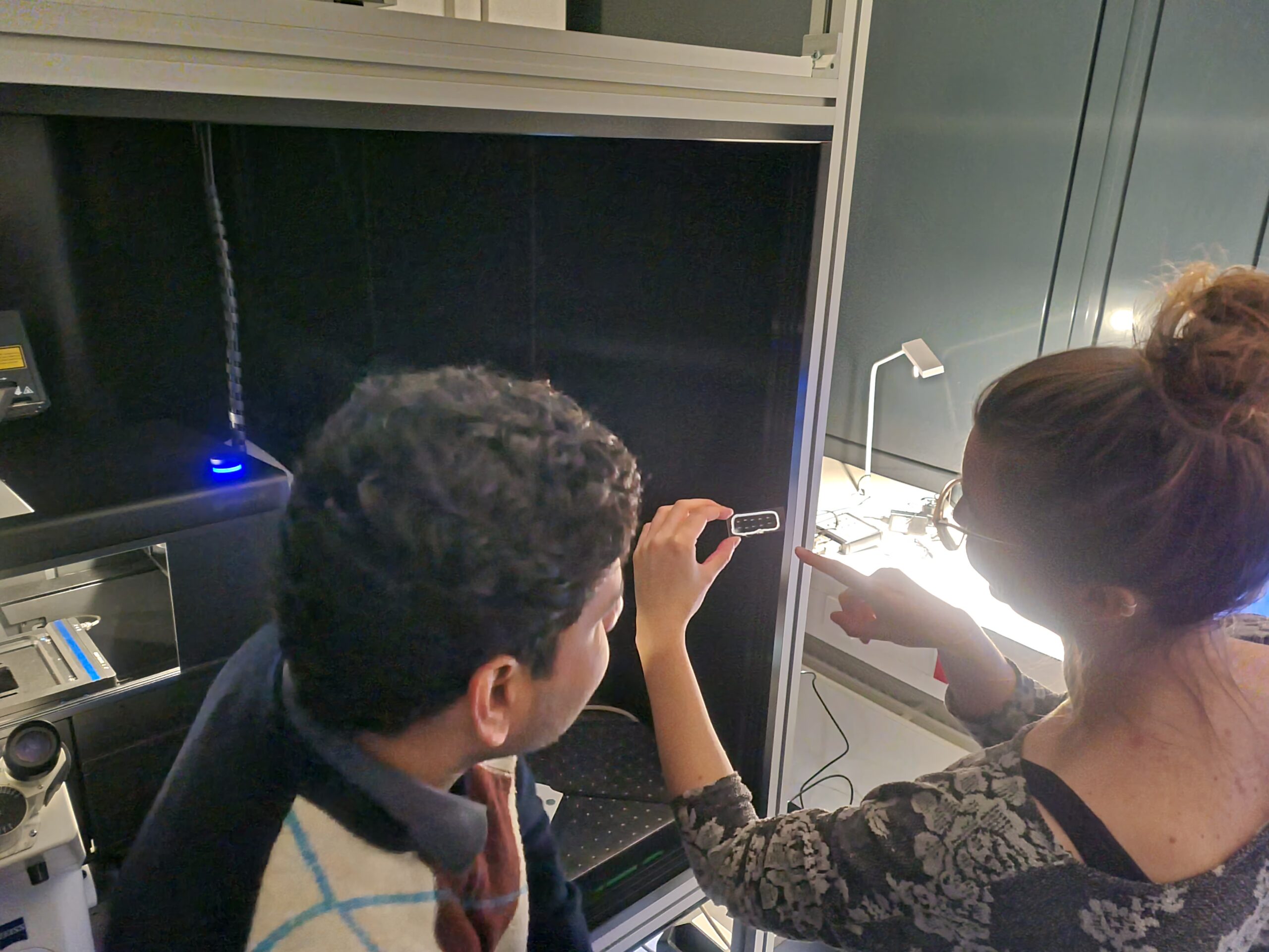
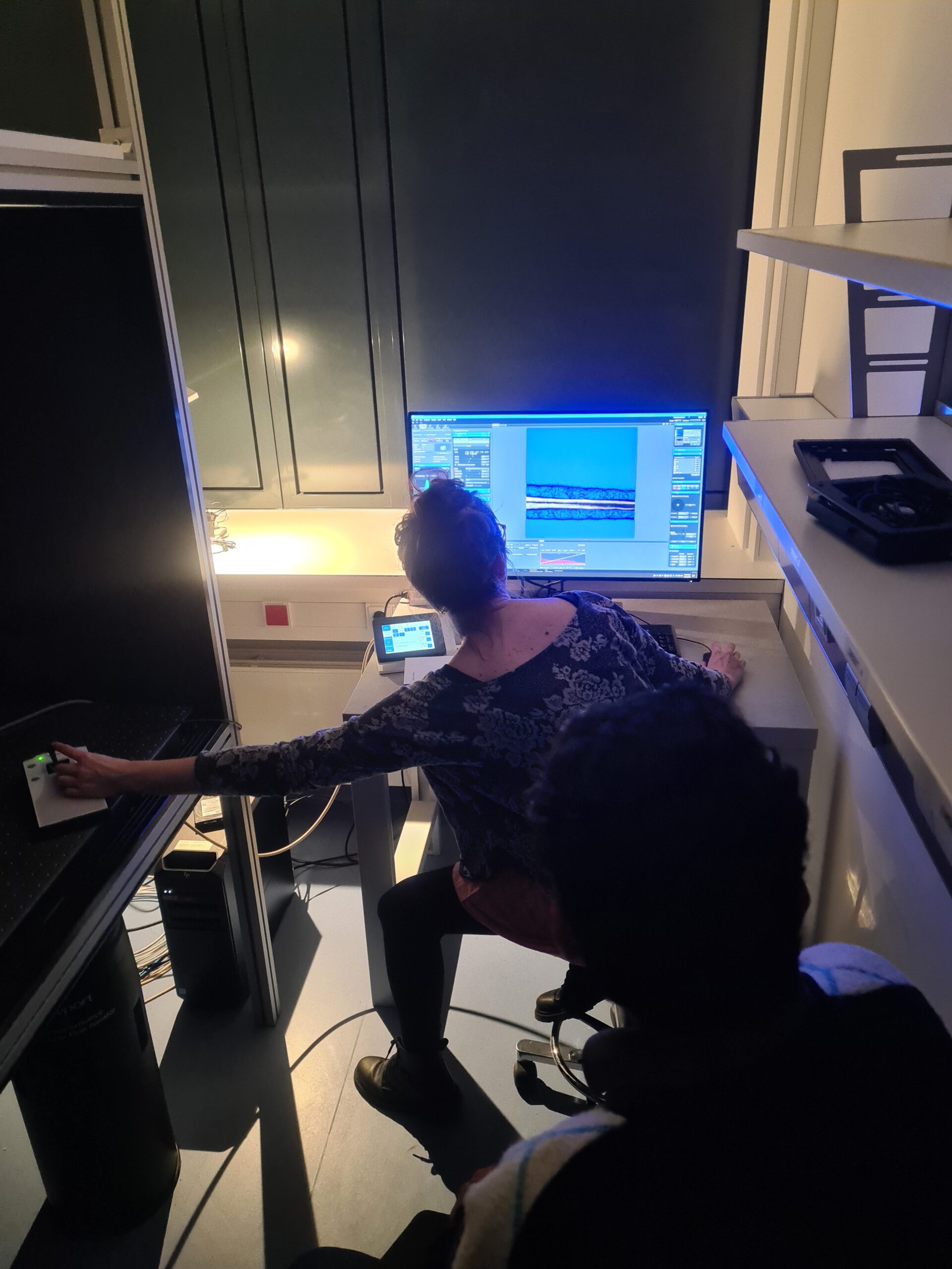
We recently wrapped up a fascinating lab shadowing with the B01 and B05 teams! During the visit, Nora John from Daniel Wehner’s lab (B05) showcased how confocal microscopy is used to image zebrafish larvae during spinal cord regeneration. This will be the starting point of a collaborative effort to characterize the morphological changes the spinal cord undergoes during healing. The findings will serve as the basis for continuum mechanics-inspired models for an in silico simulation of central nervous system regeneration in zebrafish and other species to test possible treatments for spinal cord injury with computational methods.
The collaboration with Rahul G. Ramachandran and Oskar Neumann (B01), supervised by Paul Steinmann and Silvia Budday, combines these observed biological processes with their continuum-based computational framework for spinal cord regeneration – a multidisciplinary effort that contributes to our understanding of tissue mechanics, axon growth and regenerative medicine.
Oskar Neumann, B01 & Daniel Wehner, B05